


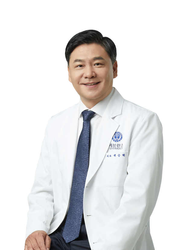
Hospital Director / Doctor of Medicine
-
Currently
- Supreme Court expert reviewer
- Vice President of the Korean Society of Colon and Proctology
- International member of the American College of Colorectal Surgery
(International Fellow, The Americal Society of Colon and Rectal Surgeons) - Vice President of Busan Surgical Society
- Expert member of Busan Public Medical Committee
- Busan Metropolitan City Autonomous Police Policy Advisory Committee Member
- Vice President of the Korean Society of Colon and Proctology Before
- Professor, Department of Surgery, Severance Hospital, Yonsei University College of Medicine (2006-2022) - Professor at Colorectal Cancer Center (2006-2014) - Director of Colorectal Cancer Center (2015-2022) - Planning Department Director (2017-2018) - Director of Medical Cooperation Center (2021-2022)
- Vice President, Korean Peritoneal Cancer Society (2021-2022)
- Special Director, Busan Surgical Society (2023)
- Announcement of survival results for stage 4 colon cancer Hi-Pac surgery for the first time in Korea (April 2022)
- Asia’s first robotic rectal cancer surgery performed (June 2006)
- Performed the world’s first robotic rectal cancer surgery in 100 cases (2007)
- National Cancer Conquest Multicenter Study (Colon Cancer) Senior Researcher (2013-2016)
- Member of the Education Committee of the Korean Surgical Society (2012-2013)
- Member of the Public Relations and Public Relations Committee of the Korean Society of Colon Proctology (2011-2013)
- Member of the Academic Committee of the Korean Society of Colon and Proctology (2014-2015)
- Member of the Planning Committee of the Korean Society of Endoscopic Laparoscopy (2012-2013)
- Member of the Editorial Committee of the Korean Society of Endoscopy and Laparoscopy (2014-2015)
- Member of the Planning Committee of the Korean Society of Surgical Infectious Diseases (2014 - 2015)
- Digestive Disorders Research Fellow, Korean Medical Association (2007)
- Colon cancer researcher at Adult Solid Tumor Research Center (2004-2006)
- Colon cancer researcher at Jangam Biochip Development Center (2004-2006)
- Member of the Inflammatory Bowel Disease Research Group of the Korean Society of Colon and Proctology (2011~)
- Korean Surgical Society Examination Committee Member (2012)
- Member of the Korean Medical Examination Committee (2013)
- Korean Society of Surgical Infectious Diseases Planning Committee (2014-2015)
- Korean Society of Colon and Proctology Inflammatory Bowel Disease Research Group (2011-2022)
- Member, Society of Surgical Oncology (SSO) (International)
- Member, American Society of Colon and Rectal Surgeons (ASCRS) (International)
- Member, Society of American Gastrointestinal Endoscopic Surgeons (SAGES) (International)
- Member, Association of Surgeons, Gastroenterologists and Oncologists (International)
- Annals of Surgical Oncology reviewer
- Disease of Colon and Rectum reviewer
- Annals of Surgery reviewer
- Official journal of the Korean Society of Endoscopic & Laparoscopic Surgeons reviewer
- Journal of the Korean Surgical Society reviewer
- World Journal of Gastroenterology reviewer
- International Journal of Advanced Robotic System Editorial Board Member
- World Journal of Gastrointestinal Endoscopy Editorial Board Member


Gastroenterology endoscopy subspecialist
- Hanyang University School of Pre-med / School of Medicine
- Specialist in Internal Medicine (Completed Internship/Residency at Hanyang University Medical Center)
- Former Fellow in Gastroenterology, Yangsan Pusan National University Hospital
- Former Clinical Professor in Gastroenterology, Yangsan Pusan National University Hospital

Family medicine specialist
- Graduated from Pusan National University College of Medicine
- Completed residency and obtained specialist certification in Family Medicine at Pusan Paik Hospital
- Fellow in Family Medicine at Pusan Paik Hospital
- Supervising Specialist in Family Medicine at Pusan Paik Hospital
- Regular Member of the Korean Society of Gastrointestinal Endoscopy
- Regular Member of the Korean Society for the Study of Obesity

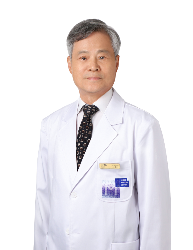
Radiologist
- Doctor of Medicine / Board Certified Radiologist
- Served as Professor at Ewha Womans University College of Medicine
- Served as Clinical Professor, Department of Health Medicine, Samsung Seoul Hospital
- Visiting Fellow, MD Anderson Cancer Center, USA
- Served as Head of the Department of Radiology at Medipium Medical Corporation's Seohyeon, Pangyo, and Dongtan Hospitals
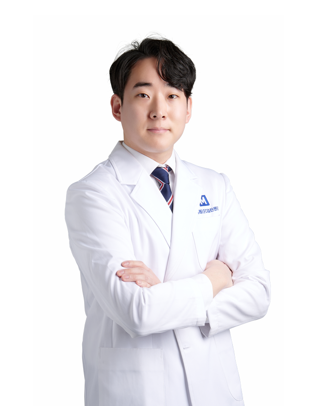
Occupational and Environmental Medicine Specialist
- Graduated from Dong-A University School of Medicine
- Completed Internship at Dong-A University Hospital
- Completed Residency in Occupational and Environmental Medicine at Dong-A University Hospital
- Certified Board Member in Occupational and Environmental Medicine
- Regular Member of the Korean Association of Occupational and Environmental Medicine
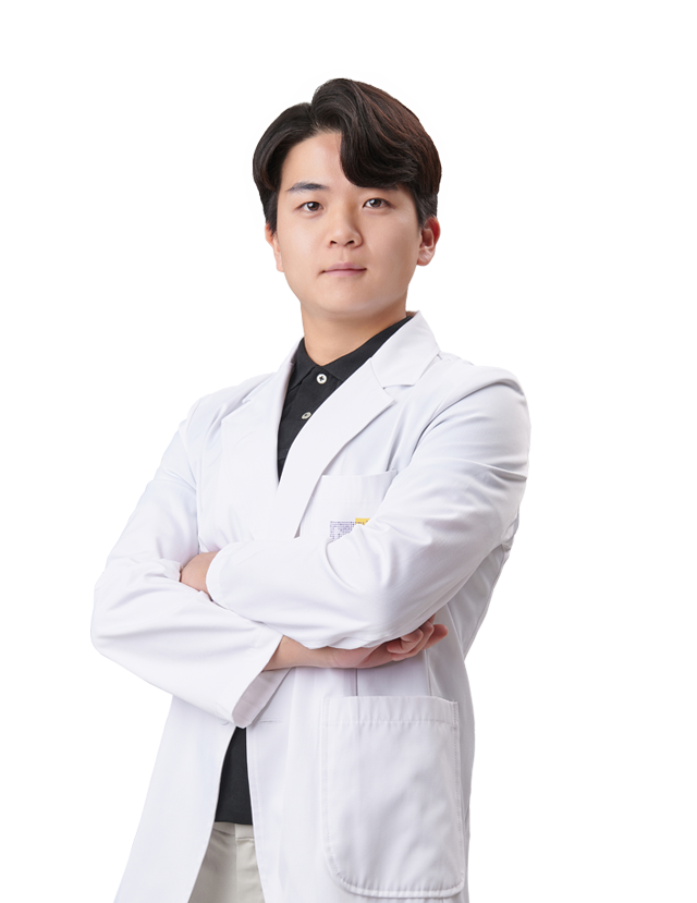
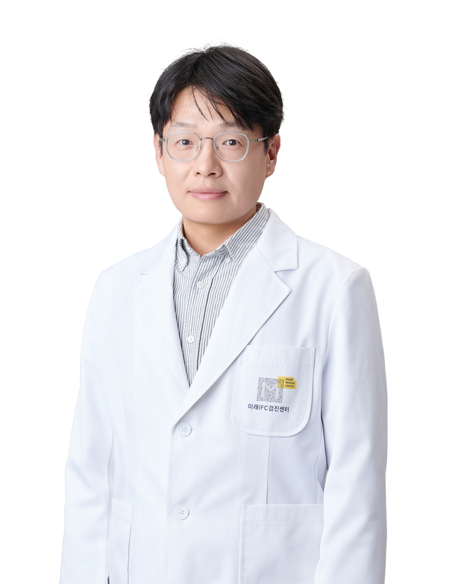
internal medicine specialist
- Chosun University College of Medicine
- Internal Medicine Specialist, Gwangju Veterans Hospital
- Former Director of Internal Medicine, Gwangju Kkori Hue Hospital
- Former Director of Internal Medicine, Gwangju Segyero Hospital
- Former Head of Internal Medicine, Busan Dongnae Health Management Association



Basic physical fitness test
- Inspection itemsDoctor consultation, height, weight, blood pressure
- Test DescriptionConsult with a specialist about subjective symptoms, diet, exercise, smoking, and drinking, and measure body measurements, obesity, and blood pressure.
Urinalysis
- Test DescriptionUrine is a waste product produced after blood is filtered by the kidneys and contains various metabolites. By testing urine, you can find out information about not only urinary tract abnormalities but also systemic endocrine/metabolic diseases. Urine is a relatively simple test that provides clues to the diagnosis of many diseases, allowing the results to be known within a short period of time without suffering.
blood test
- Inspection itemsLiver function test / Hepatitis test (Hepatitis B antibody test (HBs Ag, Anti-HBs), Hepatitis C antibody test (HCV Ab (EIA)) / Lipid metabolism / Diabetes test / Thyroid function test / Pancreas function test / Kidney function test / Tumor marker (liver cancer, colon cancer, prostate cancer, ovarian cancer) / Rheumatism inflammation test / Gout test / General blood test / Infectious disease
- Test DescriptionBlood not only plays an important role in supplying oxygen, nutrients, hormones, and nutritional substances to the cells of all organs in the body, but also functions as a defense organ against infection and transport of toxins and waste products. Therefore, changes that occur in the body lead to changes in blood composition. Various components of the blood are tested to determine the progress of the disease or the effectiveness of treatment.
Body composition test
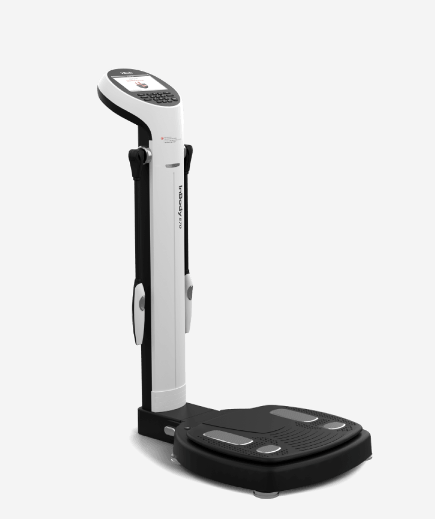
- Test DescriptionThe components that make up the body are examined. Proper management is guided by examining the ratio distribution of weight, body fat, body water, and muscle mass compared to height. A body composition test is a test that can check the degree of physical development and obesity. Through a body composition test, related diseases can be diagnosed and prevented in advance.
eye exam
- Inspection itemsVision test / fundus test / intraocular pressure test
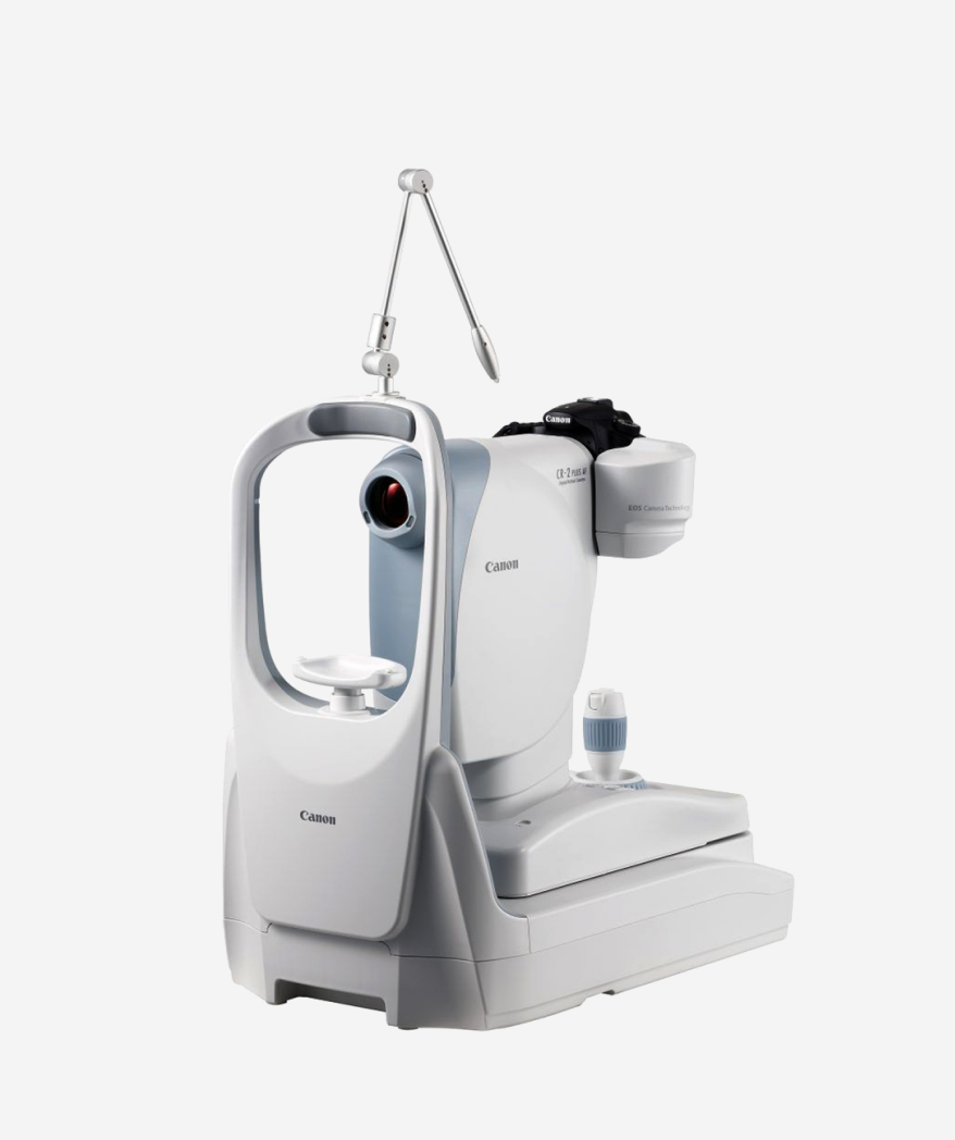
- Test DescriptionFundus audit is a test that is helpful in diagnosing eye diseases, refraction, systemic diseases, and intracranial diseases because it allows the observation of retinal blood vessels, optic nerve head, macula, and other structures in the eye using an ophthalmoscope. Observation of the eye's internal vitreous body, optic disc, retina, and choroid through a dilated pupil is not only fundamental to ophthalmological care, but is essential to its function in systemic disease states.
hearing test
- Test DescriptionA test that measures the level of hearing is called a hearing test. This is a test that measures the sensitivity of your hearing, such as whether you can hear sounds or voices well.
cardiography
- Test DescriptionIt is a test that can evaluate arrhythmias, ventricular and atrial hypertrophy, myocardial ischemia and myocardial infarction, heart disease, systemic diseases affecting the heart, effects of heart medications, abnormalities in electrolyte metabolism (especially calcium and potassium), and pacemaker function of artificial heart pacemakers.
Pulmonary function test
- Test DescriptionIn general, spirometry refers to a method of measuring the condition of the lungs by mechanically measuring the breathing process of inhaling and exhaling air. It is a test that uses normal breathing or a breathing method of inhaling as much as possible and exhaling as hard and fast as possible.
chest x-ray
- Test DescriptionIt is a test that diagnoses pneumonia, pulmonary tuberculosis, pulmonary suppuration, lung cancer, pulmonary hemorrhage, and cardiopulmonary disease (silicosis), and can also detect emphysema, pulmonary cysts, and pneumothorax. In addition, patients with valvular heart disease, congenital heart disease, and high blood pressure, as well as the presence of rib abnormalities, can be identified by the enlargement of the heart.
arteriosclerosis test
- Test DescriptionArteriosclerosis is a type of aging phenomenon in which fat, cholesterol, and neutral fat are deposited in blood vessels, narrowing the lumen of blood vessels and losing elasticity. Although there are differences in severity, it can be seen in all elderly people. By analyzing factors that can cause arteriosclerosis and conducting early tests, we can prevent arteriosclerosis and subsequent cardiovascular diseases.
abdominal ultrasound
- Test DescriptionThis is a painless test that uses ultrasound to image cross-sectional images of internal organs from various angles. It examines the liver, kidneys, gallbladder, pancreas, and spleen to diagnose liver cancer, gallbladder cancer, gallstones, pancreatic cancer, kidney stones, and kidney cancer.
gastroscopy
- Test DescriptionGastroscopy is a test method that uses an instrument to observe the digestive organs such as the esophagus, stomach, and duodenum through the oral cavity, and if there is an abnormality, it is diagnosed through a biopsy.
Gastrointestinal contrast test
- Test DescriptionGastrointestinal contrast examination is a test that allows you to observe defects, polyps, inflammatory diseases, stomach tumors, etc. by taking a contrast medium that does not penetrate gastric contrast radiography and then applying or filling the posterior wall of the lesion area. It is used for people who have an aversion to endoscopy or for mass rectal examinations, and if an abnormality is found, it is recommended to perform an endoscopy to confirm treatment. You should fast after dinner the day before the test, and it is recommended to drink plenty of water after the test to expel the contrast agent quickly.
bone density test
- Test DescriptionThe normal value of bone density varies depending on each test method, age, and gender. In other words, by measuring bone mass (bone density), we provide evidence to check for osteoporosis or bone mass loss in advance and prevent osteoporosis.
Gynecological examination (female)
- Inspection itemsCervical cancer screening
- Test DescriptionCervical cancer screening is a test in which cells from the cervix are collected using a cotton swab and then examined under a microscope. There is little pain during the test, and it tests for gynecological diseases such as cervical cancer and vaginitis.
Breast cancer screening (female)
- Test DescriptionIt takes pictures up and down, left and right, and is useful for detecting mammary tumors and mammary cysts. Mammography is very meaningful to observe suspicious signs of cancer, such as asymmetric shading and calcification. However, dense breasts require additional detailed examination using ultrasound.
Thyroid ultrasound
- Test DescriptionThyroid ultrasound is used to observe the shape, size, and nature of nodules in the thyroid gland. In particular, if there is a nodule in the thyroid gland, it is possible to determine whether the contents of the nodule are solid or cystic, and it is useful for tracking changes in the size of the nodule after treatment.
Prostate ultrasound (male)
- Test DescriptionThis is a painless test that uses ultrasound to examine the shape and size of the prostate through the lower abdomen.
Pelvic (uterine) ultrasound (female)
- Test DescriptionUterine ultrasound is a test that determines the thickness of the endometrium, uterine fibroids or adenomas, ovarian cysts, ovarian cancer, and the condition of the ovaries.
Carotid ultrasound
- Test DescriptionThis is a test that diagnoses and evaluates blood flow within the carotid artery and the condition of blood vessels using ultrasound. Diagnosis is made for partial arterial occlusion or arterial stenosis, which causes reduced blood flow to the brain.
Sleep gastroscopy
- Inspection items
- Test Description
MDCT
- Inspection itemsHead CT / Lung CT / Lumbar CT / Cervical CT
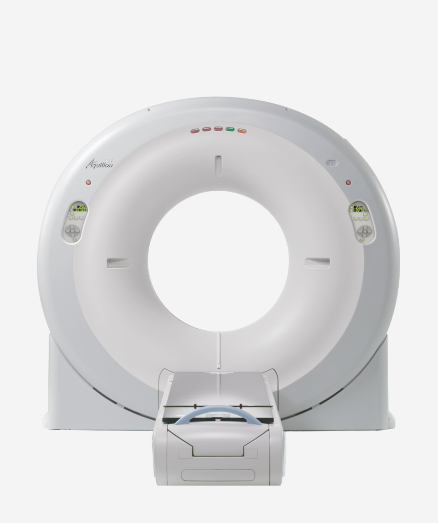
- Test DescriptionCT can image cross-sections of the human body in a short time using radiation, and is more sensitive than MRI, especially for bone microfractures, calcified lesions, and cerebral hemorrhage.
HPV virus test
- Test DescriptionHPV is a human papillomavirus and is a common virus that affects 50 to 80% of men and women who have sex at least once. It is the causative agent of reproductive cancers such as cervical cancer and vaginal cancer, and 99% of cervical cancer is caused by HPV.
echocardiogram
- Test DescriptionThis is a comfortable and safe test that uses ultrasound to observe the structure and function of the heart and identify blood flow within the heart, allowing for early diagnosis of signs of myocardial infarction or other heart diseases.
MRI scan
- Test DescriptionIt provides high-resolution images when imaging soft tissues such as muscles, cartilage, ligaments, blood vessels, and nerves that are not easily visible on CT. This is a very important test for brain (cerebral infarction, brain tumor, etc.), head and neck, and musculoskeletal diseases.
Heart (coronary artery calcification)
- Test DescriptionThis is a test that takes pictures of the coronary arteries, which are blood vessels that supply blood to the heart muscle, and estimates the degree of calcification in the coronary arteries, giving the degree of calcification a score.
MRA
- Inspection itemsThis test checks for abnormalities in the brain blood vessels and can diagnose cerebrovascular diseases such as cerebral aneurysms and vascular malformations.
Autoimmune test (CTD SCREEN)
- Inspection itemsIt is a test that screens for autoantibodies against intracellular components using the principle that various autoantibodies are detected at high frequency in the blood in autoimmune diseases. It consists of 14 antigens that are clinically meaningful in the diagnosis of connective tissue diseases and helps in early diagnosis of the disease.
Contrast-enhanced CT
- Inspection itemsContrast-enhanced CT is an imaging test that precisely evaluates the anatomical structure and hemodynamic characteristics of human organs and blood vessels by intravenously injecting an iodine-based contrast agent and then performing multi-detector computed tomography (MDCT). By administering a contrast agent, the difference in contrast enhancement between normal tissue and lesions can be clarified, allowing more accurate diagnosis of the presence and extent of tumors, inflammatory lesions, vascular abnormalities, hemorrhage, and embolism.
It is especially useful for differentiating tumors in abdominal organs such as the liver, pancreas, and kidneys, evaluating lung lesions, and diagnosing aortic disease, pulmonary embolism, and abdominal disease.
MRA test (magnetic resonance angiography)
- Inspection itemsMRA is a test that non-invasively evaluates the structure and blood flow of blood vessels using the physical principles of magnetic resonance imaging (MRI).
It does not use radiation and precisely images vascular lesions through non-contrast techniques (Time-of-Flight, Phase Contrast, etc.) using blood flow signals.
It is widely used in the diagnosis and follow-up of cerebrovascular diseases such as cerebrovascular stenosis and occlusion, aneurysm, vascular malformation, and carotid artery disease, and is also applied to the evaluation of the chest, abdomen, and lower extremity blood vessels when necessary.
Test name
- Inspection items
- Test Description
YONSEI MIRAE IFC
With Yonsei Mirae IFC Examination Center
Thanks to today
Tomorrow’s life will be different.
We are your changed
Every day is more important.

VIPs and different movement routes for men and women
High-quality facilitiescomfortable and spacious space
‘Healing checkup with art’ was implemented by arranging an ultra-large duplex structure and plan interior with a total floor area of 6,600 m².
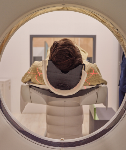
High-resolution cutting-edge imaging equipment By building
High-precision examination
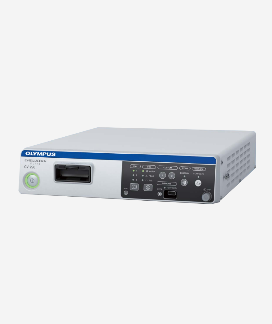
3.0T MRI / 128CH MDCT / high-resolution ultrasound equipment / digital X-ray / Olympus 290 high-definition endoscope, etc. increase the precision of examination.
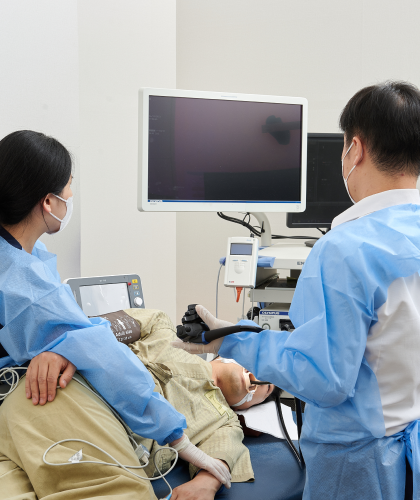
One-day, one-stop premium checkupsecond
High convenience and satisfaction by building
We have established a ‘one-day, one-stop premium checkup’ where health checkup results can be checked on the same day, linking treatment and care appropriate to the situation.

Considering each individual
Customized checkup management
Through differentiated immune tests, we provide specialized checkups that take into account not only genetic factors but also underlying diseases by age group.

Beyond diagnosis
lifelong health care
Based on the results of the examination, we provide step-by-step follow-up services for patients with symptoms, including prescription and treatment, tertiary care institution linkage service, and health information service.
Checkup reservation
Equipped with the highest specification, excellent examination equipment and professional medical staff.
A university hospital-level, state-of-the-art premium examination center your Lifelong health guardian I'll go.
CS CENTER
Do you have any more questions?
Please call the main number and we will be happy to assist you.


CONTACT
Clinic hours
133 Jeonpo-daero, Nam-gu, Busan
Go to directions


- Early detection of stroke, cerebral hemorrhage, and dementia is important.
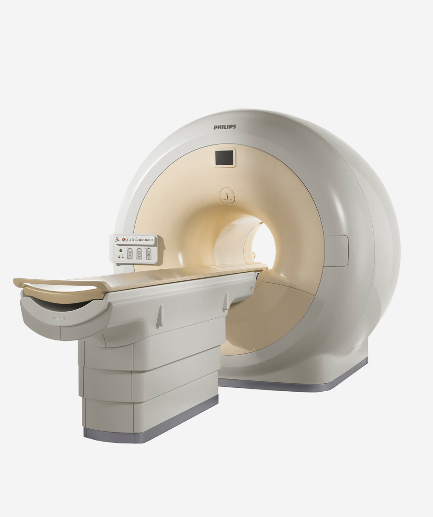
- The T in front of MRI indicates the magnetic field strength in Tesla units. The larger the number of T, the higher the resolution. 3.0T MRI has more than twice the resolution and faster imaging speed than 1.5T.
- The higher the resolution of MRI, the clearer the image results can be obtained. Small lesions, nerves, and blood vessels that could not be identified with existing low-resolution MRI Diseases can be accurately diagnosed through 3.0T MRI.

- ▣ Equipped with Philips’ proprietary MR technology!
- ▣ Resolution more than 2 times higher than existing 1.5T MRI
- ▣ Utilizes magnetic fields that are harmless to the human body

- 3.0T MRI (Magnetic Resonance Imaging) uses a superconducting magnet to generate a magnetic field and remove hydrogen atoms in the body. This is a device that obtains images of the characteristics of the human body through resonance.
- About soft tissues such as the brain, spine, musculoskeletal system, liver, pancreatobiliary duct, pelvis, and breast. Precise images are captured without the use of contrast media.
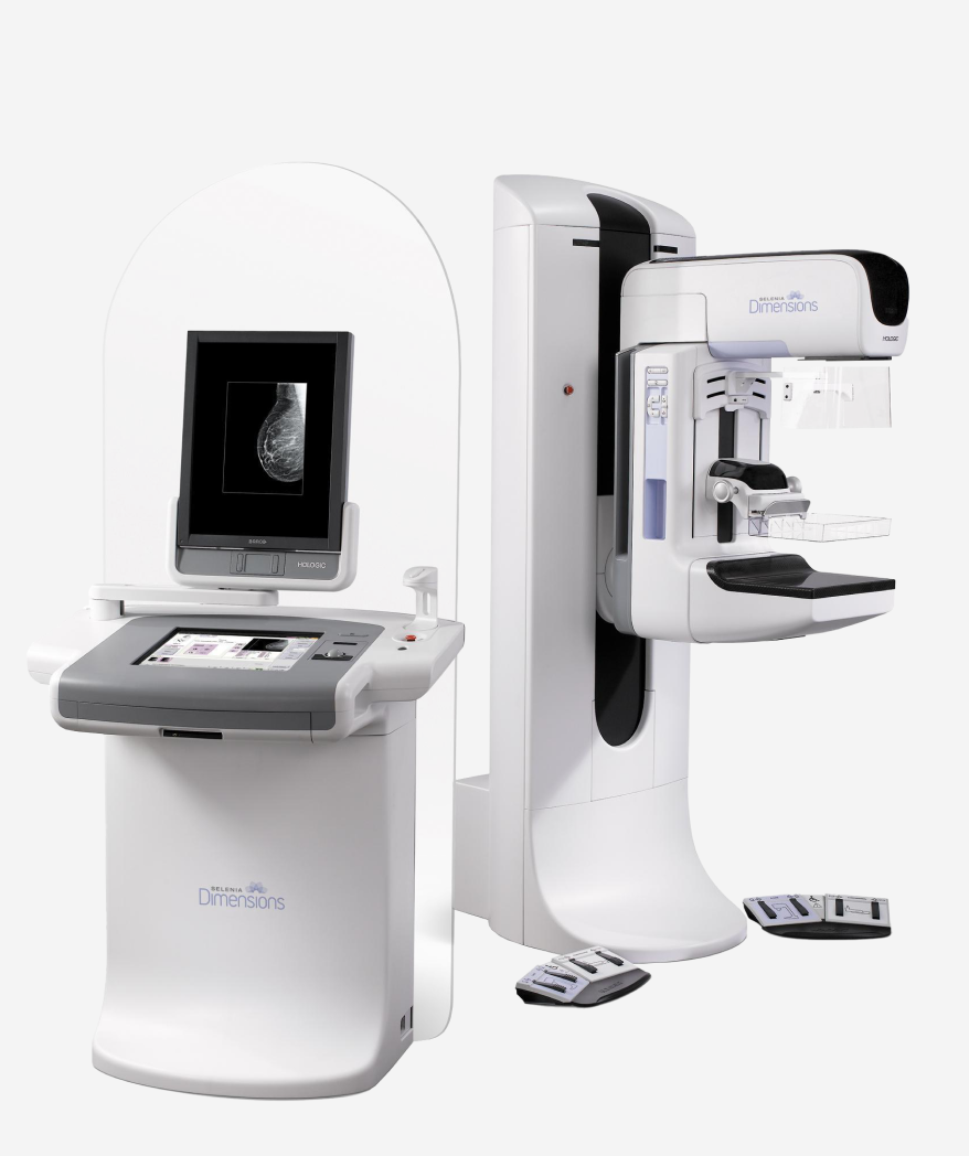
- Early diagnosis of breast cancer with artificial intelligence (AI) breast cancer imaging equipment!
- Selenia Dimensions from Hologic, USA, is the first in the world to be approved by the FDA. Through artificial intelligence (AI)-based computer-assisted diagnosis approved by MFDS We provide early diagnosis of breast cancer using mammography, the optimal medical equipment for cancer diagnosis.

- ▣ The world’s best mammography equipment approved by FDA and MFDS
- ▣ Minimize radiation exposure (1.2mGy)
- ▣ Precise diagnosis with 13.6 million pixel high-definition images
- ▣ Use of ergonomic breast compression paddle (FAST Paddle)

- 13.4 million won for testing equipment used to diagnose breast cancer early. Precise diagnosis is possible through high-definition images of pixels.
- Additionally, it automatically adjusts the optimal radiation dose to minimize unnecessary radiation exposure.
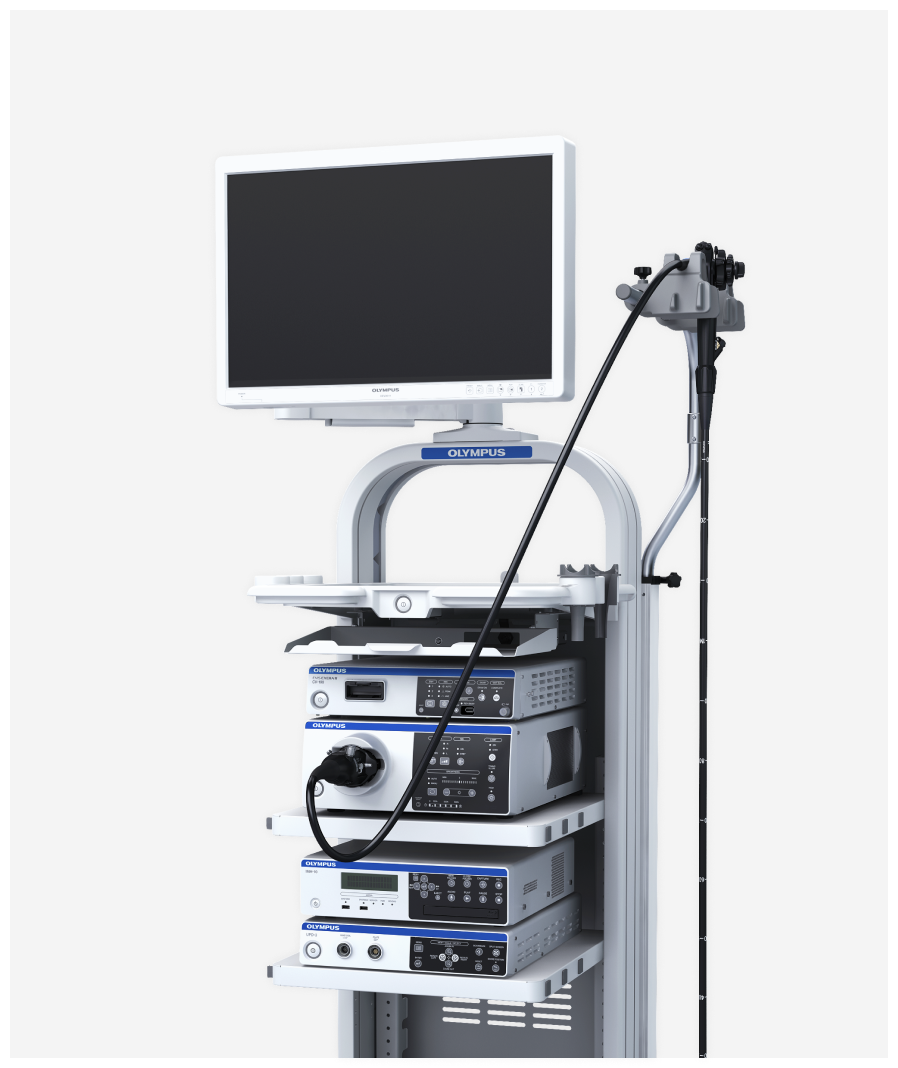
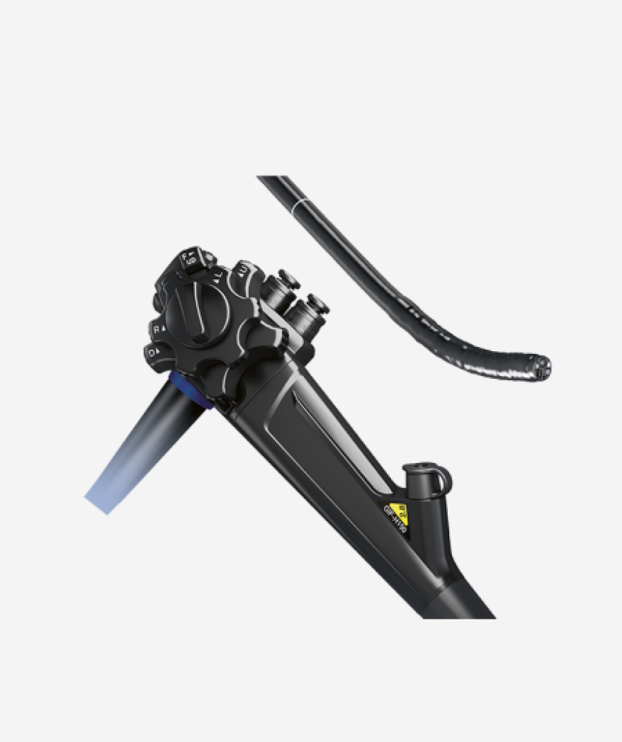
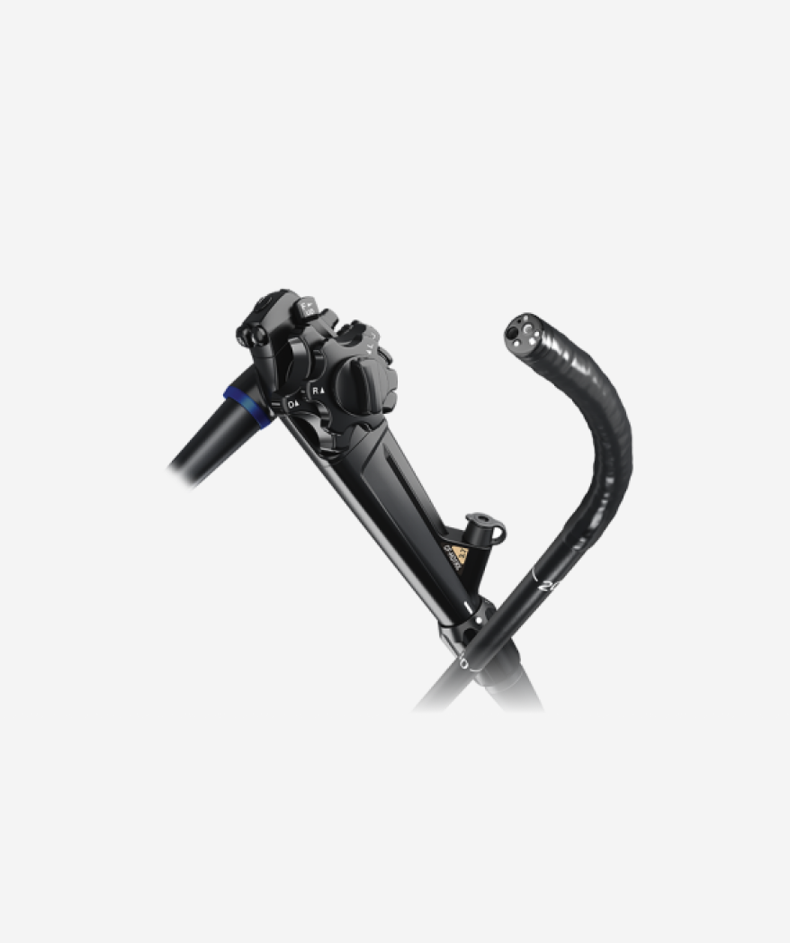
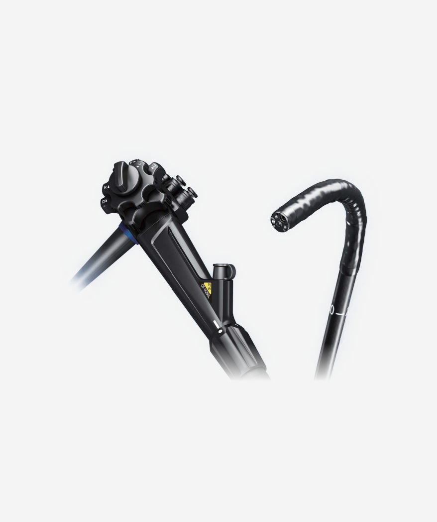
- The highest specifications among existing endoscopy equipment
- Olympus' 290 endoscope model, known as the best endoscope on the market to date, is currently being introduced and operated in ALL.

- Accurate diagnosis with high-quality medical equipment
- Colonoscopy can detect colorectal cancer, colon polyps, various inflammatory bowel diseases, and ulcerative diseases. Colon cancer is a small polyp that grows larger and turns into cancer, so early colonoscopy, even if there are no symptoms, is the only way to prevent colon cancer from developing.
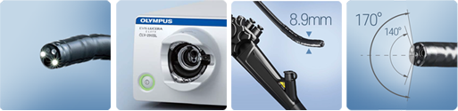
- High stomach and colon diagnosis rate
- New optical design increases the number of light guide fibers Sensitivity and brightness have been improved to detect fine details that are difficult to see with the naked eye. Small and unclear early cancer tissues can be easily distinguished.
- Pain relief function during colon examination
- The thin and flexible endoscope is convenient for insertion. Conventional colonoscopy due to smooth entry at the bend area Patients experience less pain than during the test.
- Bright and clear high-resolution HD video
- Difficulty-locating mucosal areas such as stomach and colon cancer, and microvascular By applying the latest optical technology, you can see in detail like a palm print. You can receive a quick and accurate diagnosis.
- Accurate diagnosis with wider viewing angle
- Due to the wider viewing angle of 170。 Wide angle Accurate diagnosis is possible without missing the surroundings.
- This video gastroscopy equipment allows for high-resolution observation with a thin insertion section of 8.9mm in diameter and excellent HDTV resolution. Additionally, the banding portion and curvature of the tip of the endoscope are short, allowing easier access to the esophagus and gastric angle.
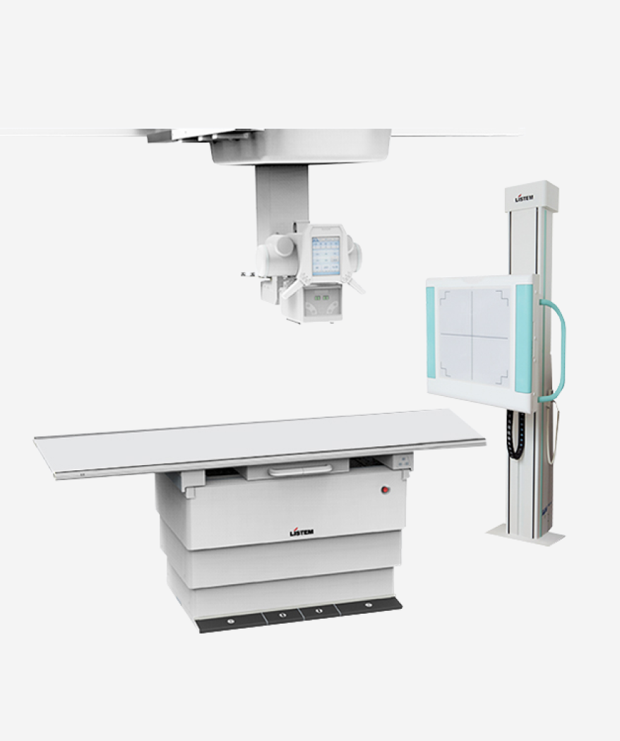
- It is a device that photographs the internal structures of the human body using ‘gamma rays’, a radioactive substance. It is commonly used to diagnose abnormalities in the lungs and bones, and is also used to diagnose various traumatic diseases including lung disease and fractures, degenerative changes in the spine and musculoskeletal system, inflammation, and tumors.
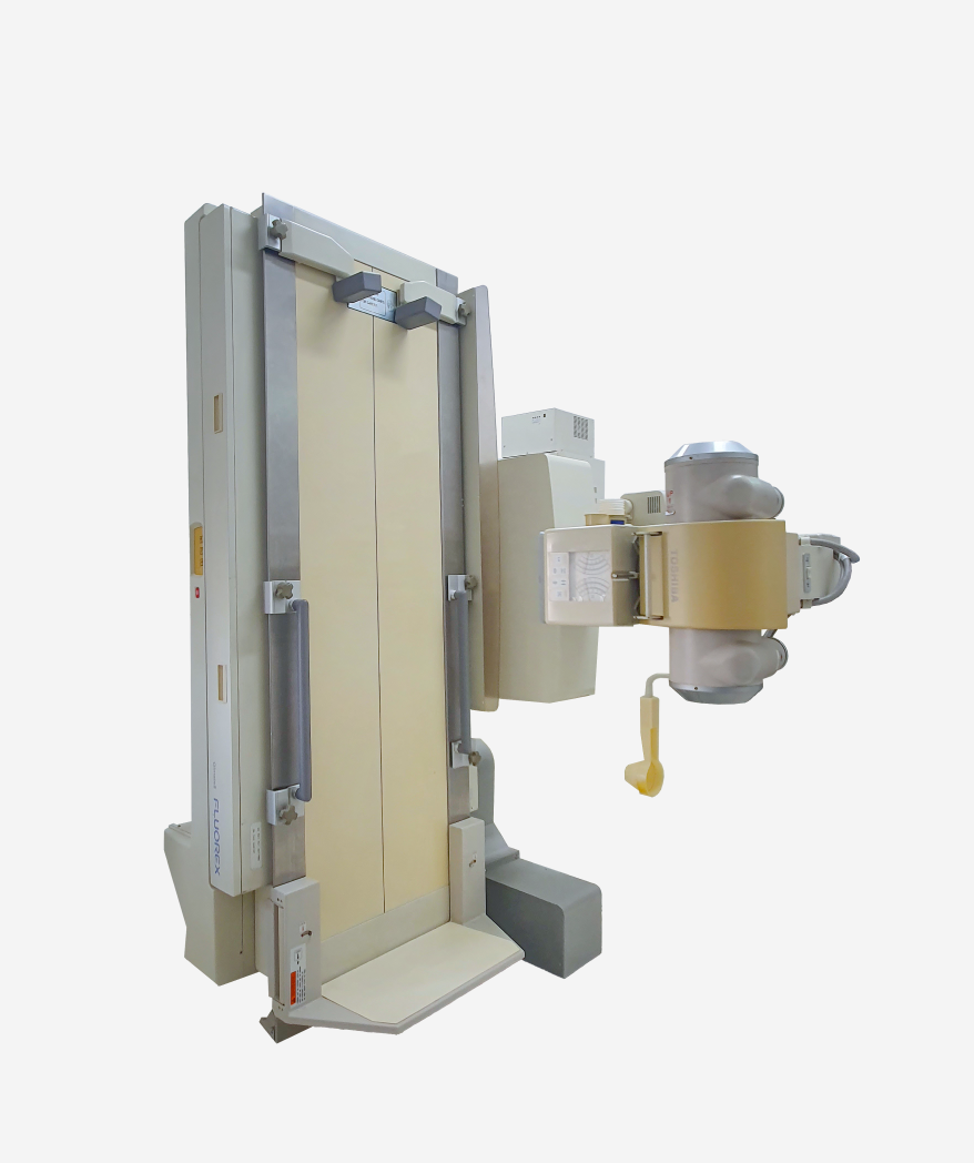
- This is equipment for an indirect examination method that observes abnormalities and functional abnormalities of the esophagus, stomach, and duodenum by taking pictures after drinking a contrast medium such as barium suspension or iodine agent that does not transmit well to X-rays. There is less risk or discomfort, but it is less accurate than a gastroscopy, so a gastroscopy may be necessary if abnormal findings are found.
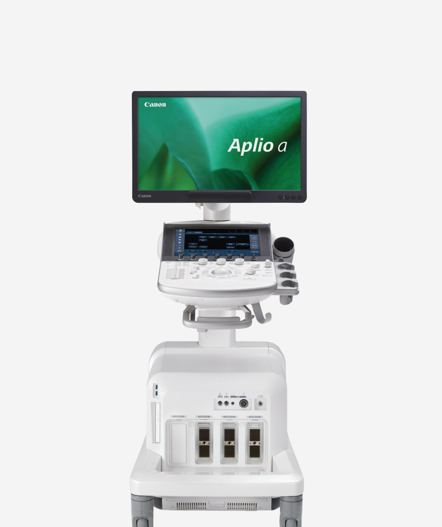
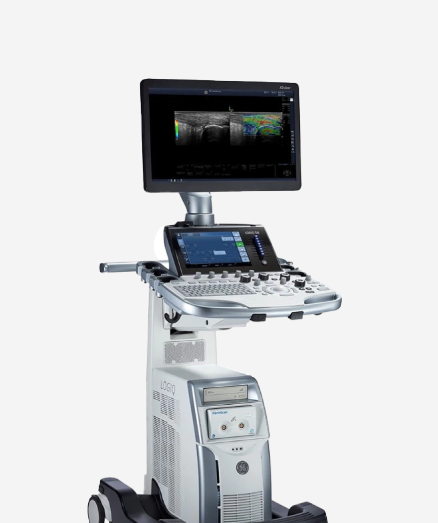
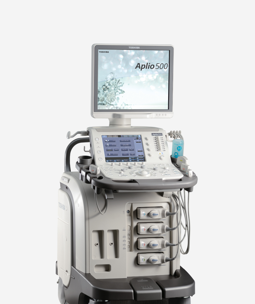
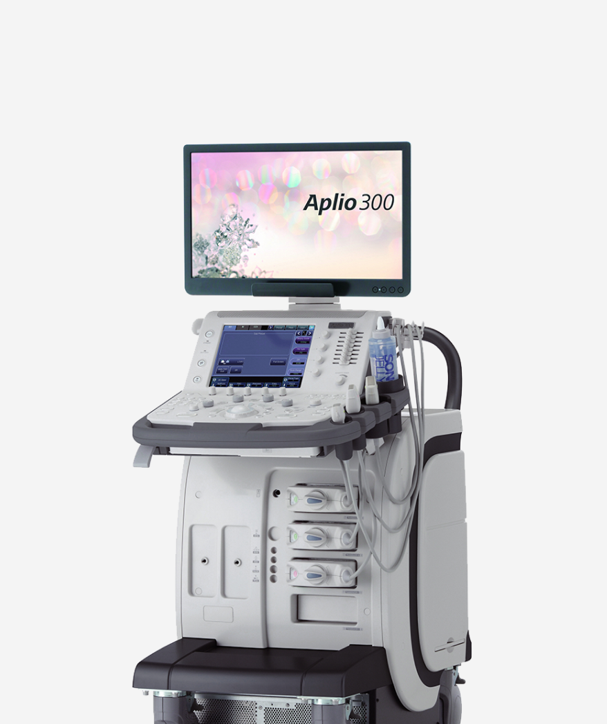
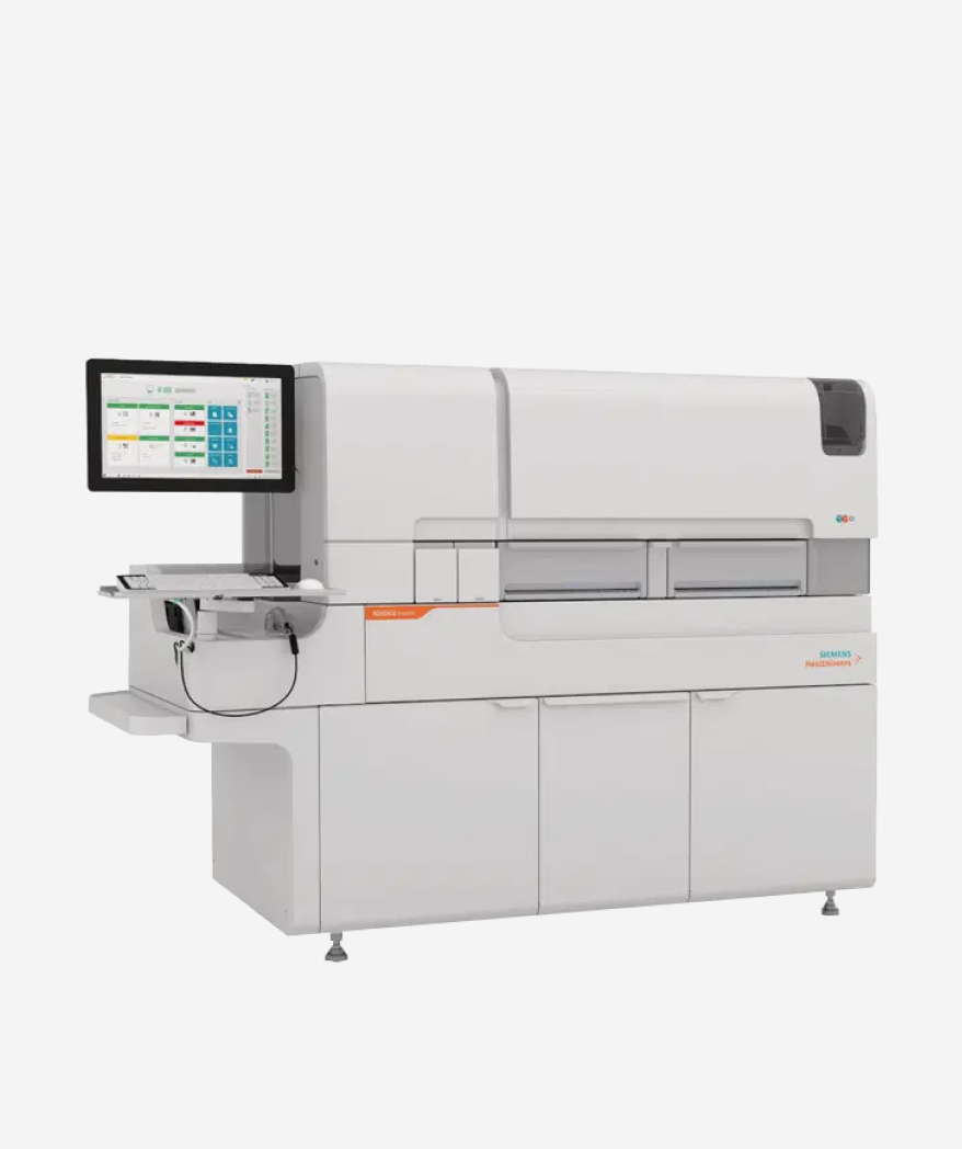
- It is an integrated biochemistry and immunology test solution that can be operated independently, and operates biochemistry and immunology tests with one device. It has the ability to process 1,000 biochemical tests and 1,120 immunological tests per hour, and can manage all data, patients, calibration, and quality control results with the efficiency of the operating software.
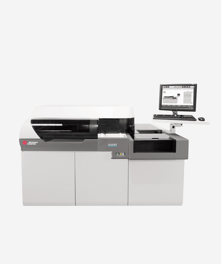
- By measuring the concentration of various substances in the blood, various enzymes related to the liver are used as indicators to diagnose liver disease and see the effectiveness of treatment. When blood sugar level is above the standard level, diabetes can be provisionally diagnosed, and jaundice and changes in kidney function can also be checked.
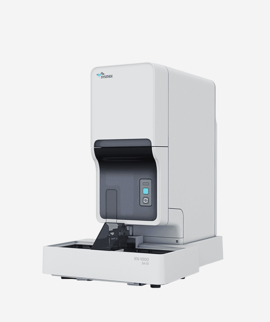
- This is a testing equipment needed to diagnose various blood diseases, such as measuring the number of blood cells in the blood or testing related to bleeding and blood clots. A processing speed of up to 100 samples per hour can be achieved. Abnormal cells are detected with high sensitivity, and measurement accuracy is improved to enable accurate measurement.
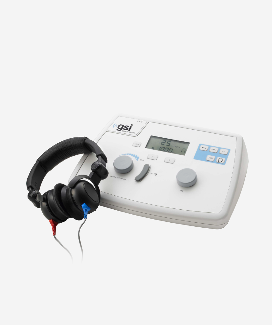
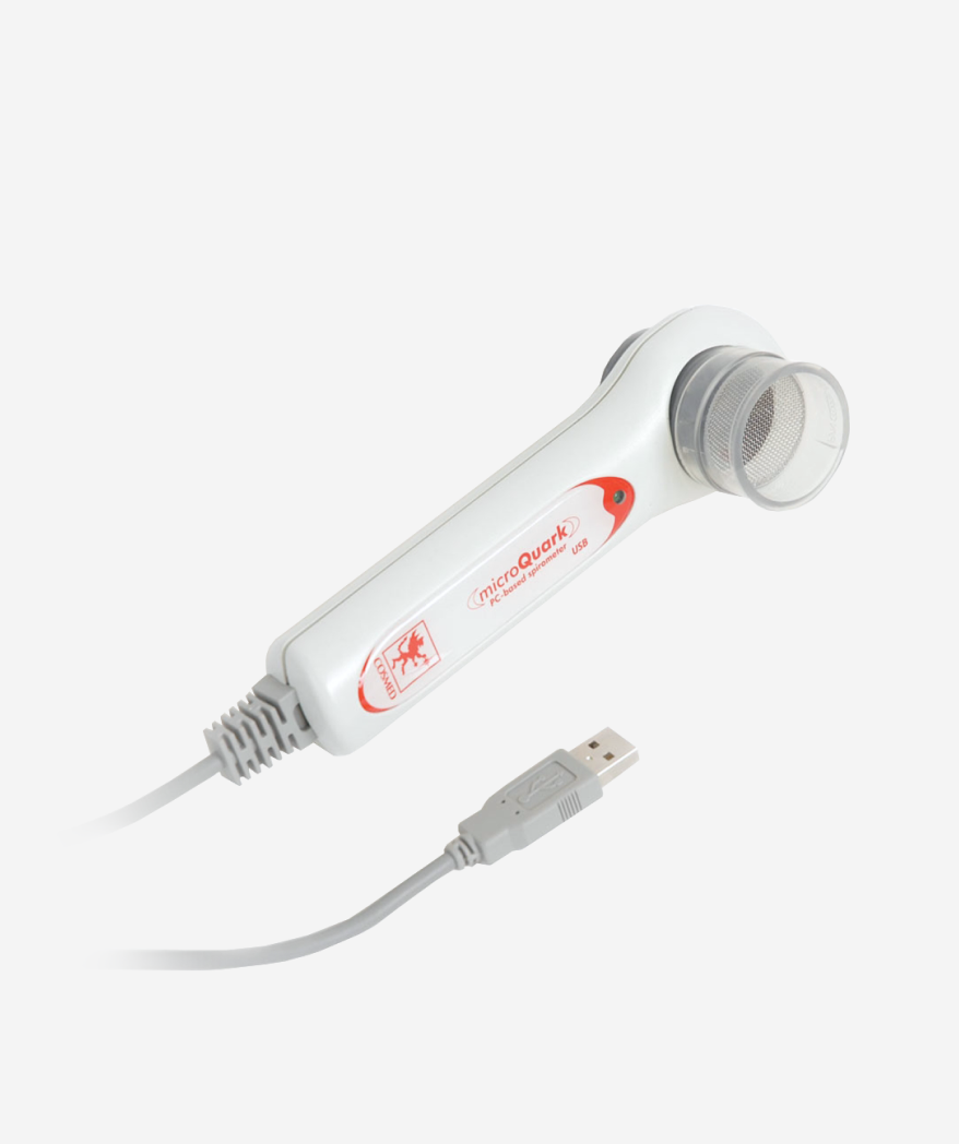
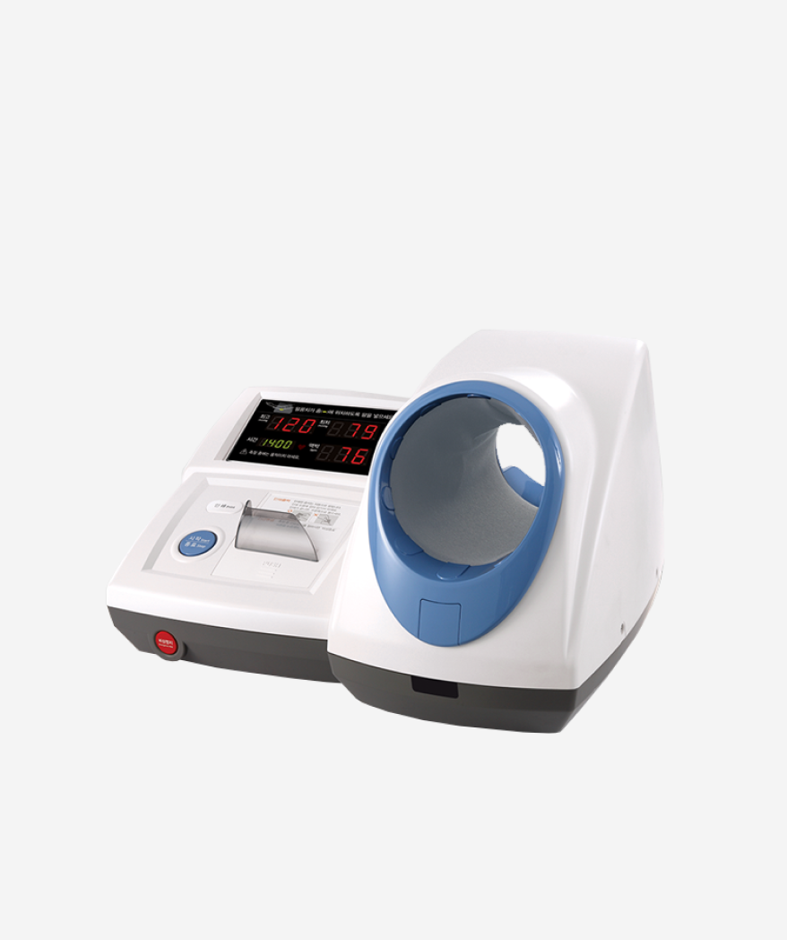
- It is a test equipment that measures the pressure exerted on the walls of blood vessels by the blood flowing along the blood vessels. The unit of blood pressure is mmHg. Normal blood pressure is around 120-130 mmHg systolic and 80-85 mm Hg diastolic. If it rises above a certain level, it is diagnosed as high blood pressure, and if it falls below a certain level, it is diagnosed as low blood pressure.
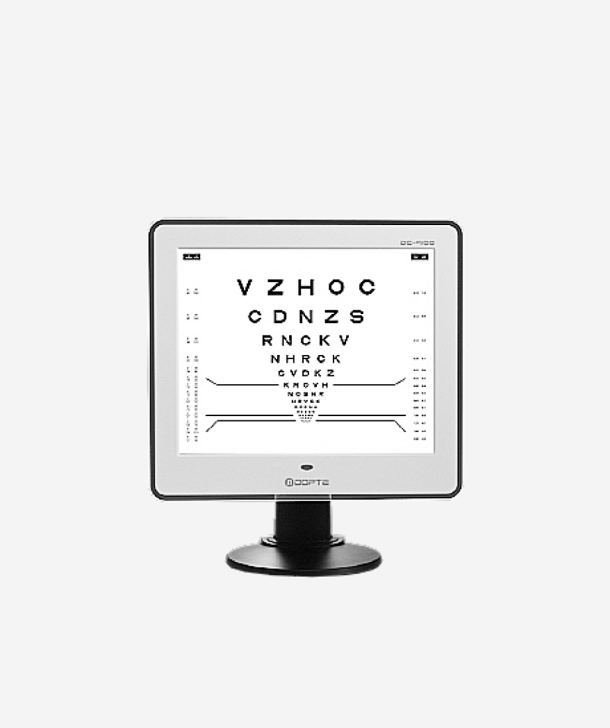
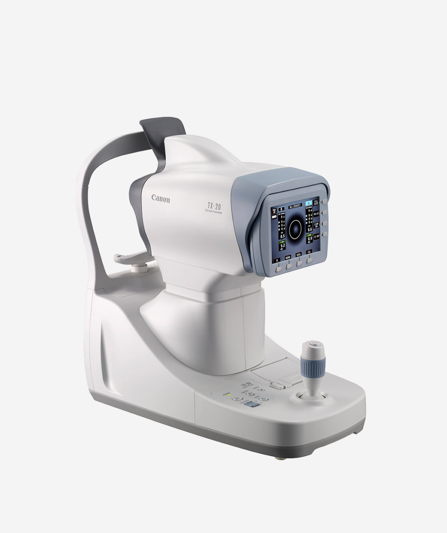
- Intraocular pressure or intraocular pressure refers to the fluid pressure inside the eye, and is a device that measures intraocular pressure. An intraocular pressure between 10 and 20 mmHg is considered normal, and ocular hypertension is considered the most important risk factor for glaucoma.
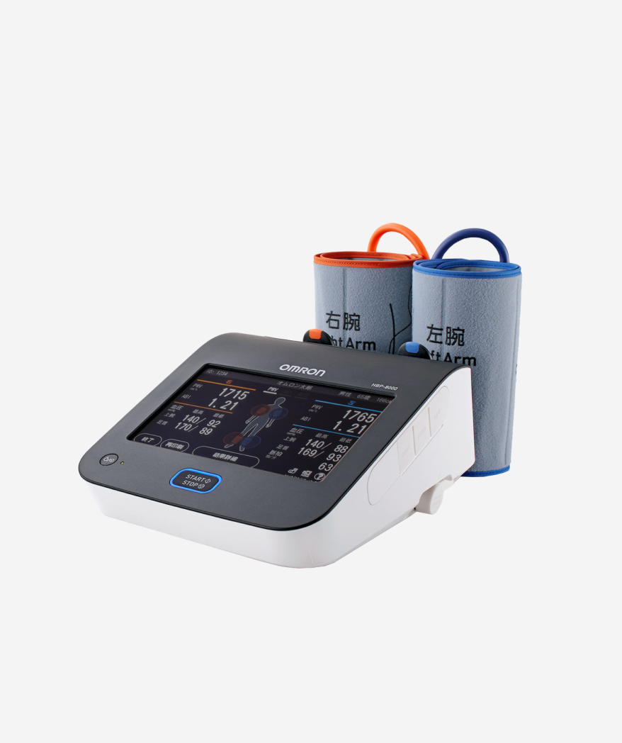
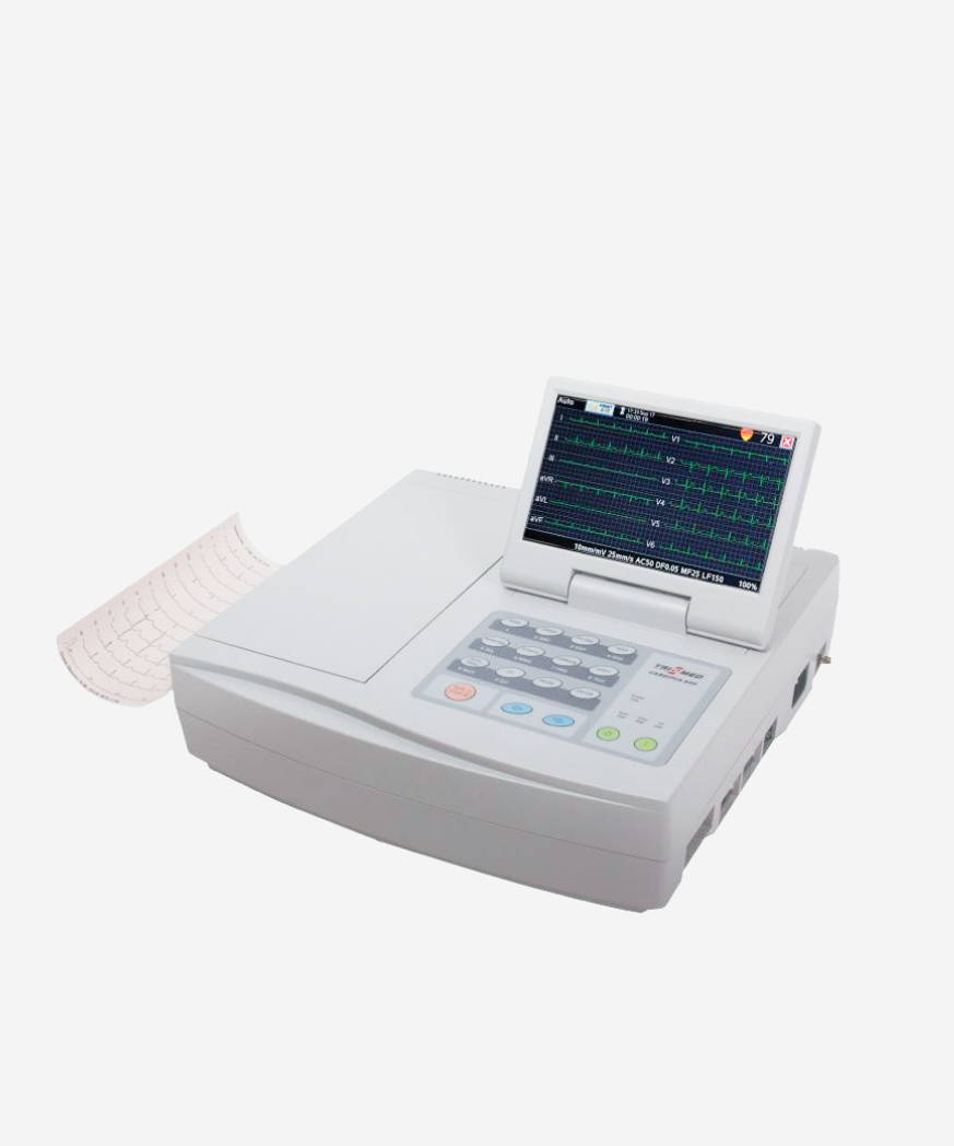
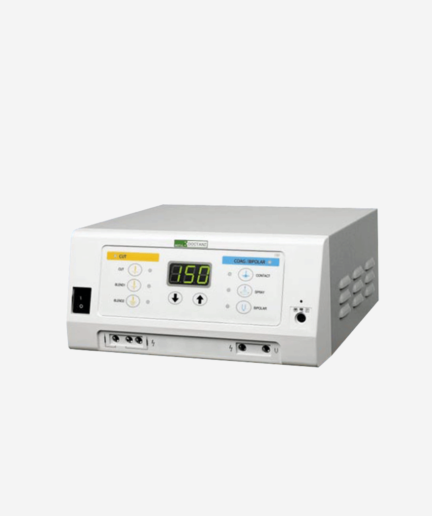
- The endoscopic Bovie is an electrosurgical device that passes high-frequency current through the body and generates heat locally to cut and coagulate during polypectomy. Safe surgery is possible by using the principle of localized vaporization, drying, coagulation, waves, and heat generation when high-frequency current within a specific range is used.
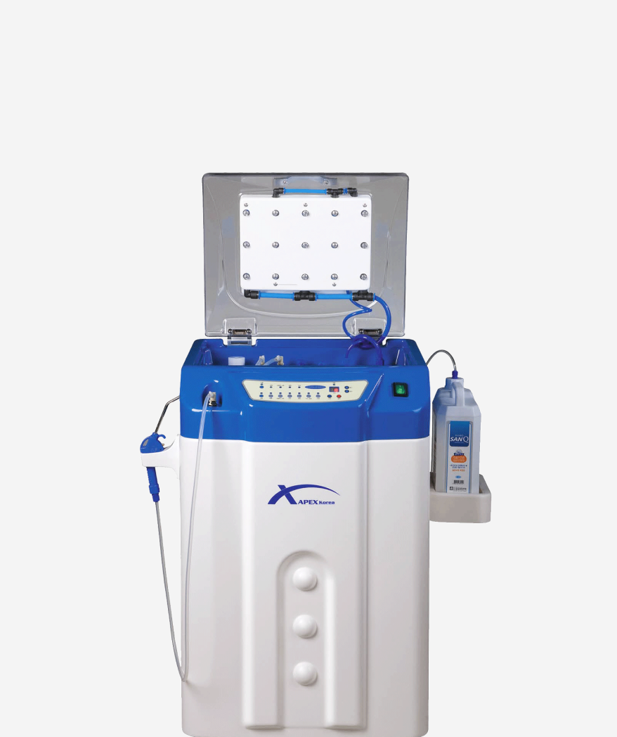
- This equipment sterilizes the scope after an endoscopy. It has an excellent cleaning effect through downward water spray using 9 cleaning nozzles. It produces high-concentration electrolytic acid water of over 1,000ppm and has a sterilizing effect through one-time use. The disinfectant is non-toxic and has environmentally friendly features.
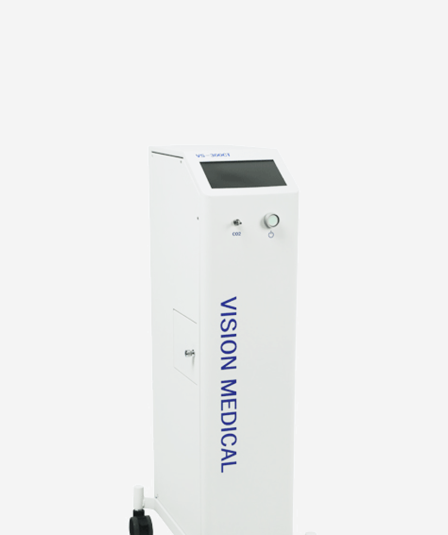
- This is a device that injects CO2 gas into the human body through an endoscope. It can prevent complications caused by gastric bypass that may occur during pancreatic surgery, and allows for a safe and comfortable examination by eliminating the pain caused by abdominal distension that occurs during colonoscopy.
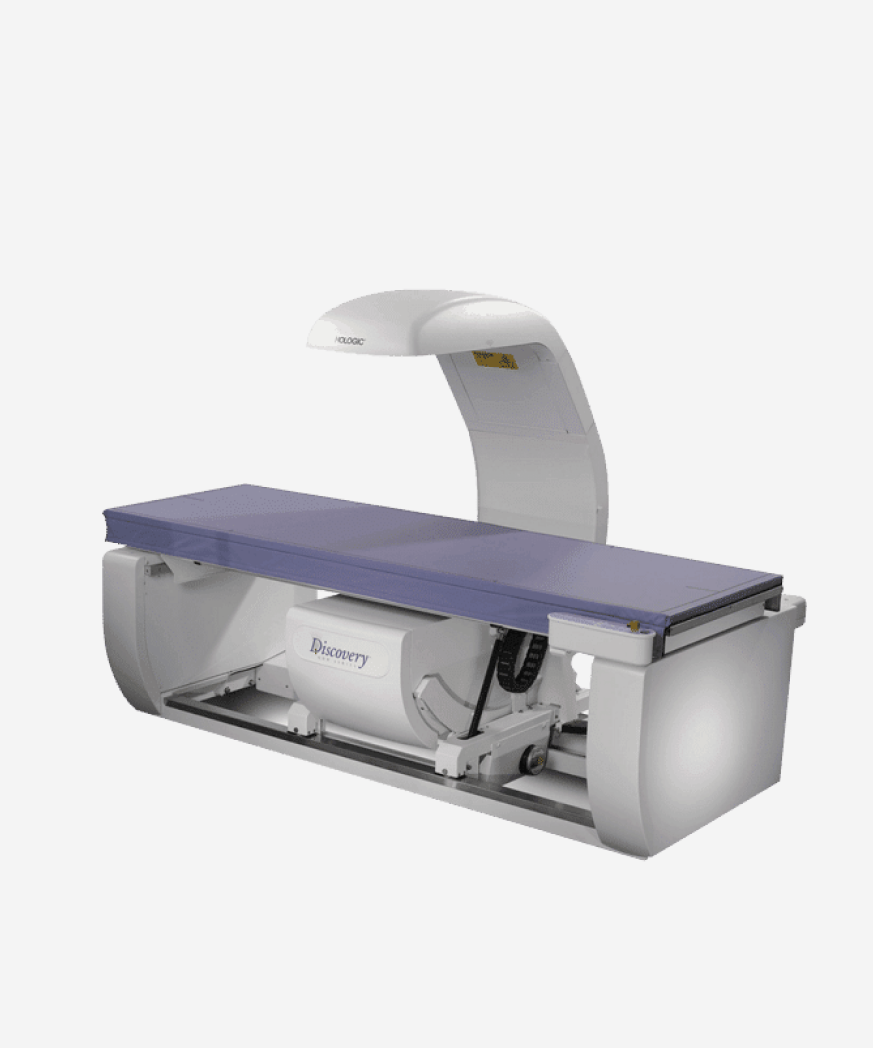
- This equipment is used to diagnose and treat osteoporosis at an early stage. It can image the front, sides, spine, wrist, etc. using an X-ray type bone density measuring device. It has high precision, receives less radiation exposure than x-ray photography, and moves automatically according to the required area.